The Cellular Gridlock: Resolving the Randle Cycle, Nutrient Competition, and Mitochondrial Inflexibility
Oct 26, 2026
Stabilize Your Energy with Evidence-Based Nutrition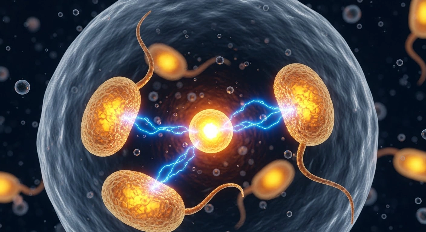
Author: Science Communication Team, Vital Life Choice LTD
Category: Upstream Metabolic Science & Chronic Disease Prevention
Reading Time: 7 Minutes
Introduction: The Myth of the "Slow Metabolism"
A ubiquitous complaint in modern health consultations across Australia is the feeling of a "sluggish metabolism"—manifested as stubborn visceral fat, unshakeable mid-afternoon brain fog, and intense lethargy following meals. Standard public discourse frequently frames this as an inevitable consequence of aging or an inherited genetic disadvantage.
At a cellular level, however, what patients experience is rarely a quantitative deficiency in metabolic rate; rather, it is metabolic inflexibility—the inability of cellular mitochondria to alternate smoothly between oxidizing glucose and oxidizing fatty acids based on physiological demand.
When standard Western dietary habits consistently combine ultra-processed carbohydrates with high-temperature oxidized seed oils (e.g., pastries, loaded takeout burgers, deep-fried chips), the cell's metabolic machinery enters a biochemical gridlock first described in 1963 by Sir Philip Randle: The Glucose-Fatty Acid Cycle (The Randle Cycle).
1. The Biochemistry of the Randle Cycle: A Defense Mechanism Turned Pathology
Under ancestral physiological conditions, the Randle cycle serves an essential protective purpose: during periods of prolonged fasting or endurance exertion, elevated rates of fatty acid beta-oxidation generate high concentrations of mitochondrial acetyl-CoA, NADH, and citrate.
Citrate Translocation: Mitochondrial citrate exits into the cytosol and allosterically inhibits phosphofructokinase-1 (PFK-1), the rate-limiting enzyme of glycolysis.
PDH Inactivation: Accumulated mitochondrial acetyl-CoA and NADH activate pyruvate dehydrogenase kinase (PDK), phosphorylating and shutting down the pyruvate dehydrogenase (PDH) complex.
This biochemical brake preserves scarce glucose for the central nervous system by forcing skeletal muscle to run exclusively on lipid substrates.
In the modern food environment, however, this cycle is weaponized against cellular health. When an individual consumes a meal presenting a dual excess of rapid-digesting starches and free fatty acids, both substrates arrive simultaneously at the inner mitochondrial membrane. The resulting collision over-saturates the mitochondrial electron transport chain (ETC), dramatically elevating intracellular reactive oxygen species (ROS), paralyzing PDH activity, and halting downstream glucose oxidation. Trapped intracellular glucose metabolites are subsequently diverted into toxic lipid intermediates—specifically diacylglycerols (DAGs) and ceramides—which directly phosphorylate serine residues on the Insulin Receptor Substrate-1 (IRS-1), severing downstream insulin signaling.
2. Mitochondrial Inflexibility & The Dawn Phenomenon
When skeletal muscle myocytes and hepatocytes remain locked in this competitive state:
Impaired Exercise Recovery: Muscle tissue fails to upregulate glycogen replenishment post-exercise because glycolytic intermediates cannot access the citric acid cycle.
Persistent Hepatic Gluconeogenesis: The liver loses the regulatory capacity to perceive postprandial insulin spikes. Even during overnight fasting, unmetered fatty acid flux into the liver keeps hepatic pyruvate carboxylase active, driving continuous endogenous glucose production—a primary mechanism underlying elevated morning fasting glucose (the exaggerated Dawn Phenomenon).
Beta-Cell Exhaustion: To overcome this persistent intracellular block at the IRS-1 node, pancreatic beta-cells are forced to sustain hyper-secretion of insulin around the clock, accelerating beta-cell de-differentiation and exhaustion.
3. Clinical Evidence: Substrate Clearance Following Whole-Food Matrix Swaps
4. Therapeutic Architecture: Restoring Cellular Fuel Flexibility
Resolving cellular gridlock requires removing the simultaneous flood of high-velocity glucose and inflammatory lipids, replacing it with phased, whole-food nutrient delivery:
Separating Hyper-Concentrated Substrates: Avoid combining isolated liquid starches/sugars with deep-fried fats (the fast-food triad). By utilizing slow-release, intact carbohydrates (such as wholemeal Spelt, lentils, quinoa, and soba), portal glucose enters circulation in a metered stream that does not overwhelm mitochondrial oxidation limits.
Leveraging AMPK Activation via Muscle Contraction: A brisk 10-to-15 minute walk immediately following meals initiates non-insulin-dependent GLUT4 translocation through the AMP-activated protein kinase (AMPK)pathway, drawing excess glucose into myocytes without requiring elevated insulin cascades.
Prioritizing Polyphenol-Rich Plant Lipids: Incorporating cold-pressed extra virgin olive oil, raw walnuts, and pumpkin seeds delivers monounsaturated and omega-3 fatty acids that enhance mitochondrial membrane fluidity and promote fatty acid transport into the mitochondria via carnitine palmitoyltransferase-1 (CPT-1) without causing DAG-induced insulin resistance.
At Vital Life Choice LTD, our community programs and clinical educational initiatives across South Australia focus on de-mystifying internal endocrinology. Metabolic flexibility is not an unchangeable fate—it is the biological return to clean, synchronized cellular respiration fostered by real, whole-food choices.
Disclaimer: This educational health publication is provided by Vital Life Choice LTD strictly for public health awareness and evidence-based lifestyle literacy. It is not intended as individual medical diagnosis, clinical management, or direct dietary prescription. Please consult a qualified medical or healthcare professional regarding personal metabolic conditions.